Discrimination and Size Measurement of Viruses in a Mixture
Nano-sized biological agents and pathogens such as viruses are known to be responsible for a wide variety of human disease such as the flu, AIDS and herpes, and have been used as biowarfare agents. Viral size plays a substantial role in transmission dynamics, disease outbreaks and outcomes. Rapid and accurate detection and characterization of single viral particles has become increasingly important for virology research, disease diagnosis and treatment, and biotechnology applications.

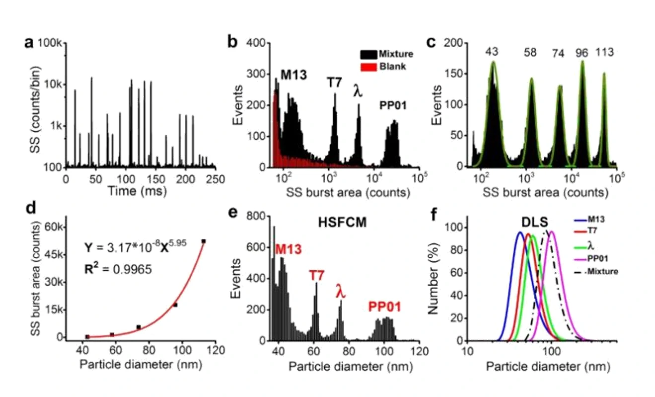
Transmission electron microscopy (TEM) has historically been the method of choice for determining the size and morphology of single viruses. However, the tedious process of sample preparation and image analysis, along with the high cost, prevents its routine use. Here, a standard calibration curve is constructed using monodisperse silica nanospheres of five different diameters ranging from 43-113 nm. The SS burst areas of silica nanoparticles are plotted as a function of the diameters determined by TEM, and the SS burst area of every single virus is then converted to the corresponding particle size.

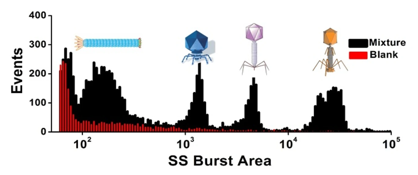
Figure 1. Differentiation and size measurement of different virus types in a mixture.
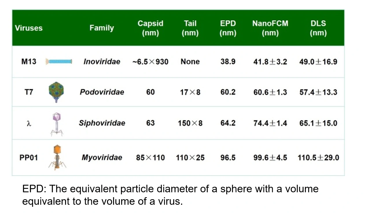
Table 1. Accuracy and precision comparison among the Flow NanoAnalyzer, DLS, and TEM for virus size measurement.

Compared with the hydrodynamic diameters measured by DLS, the optical diameters measured by the Flow NanoAnalyzer agree well with the equivalent particle diameters derived from their structural dimensions measured by TEM. This approach provides greater measurement precision in comparison to DLS.
Angew. Chem. Int. Edit., 2016, 55(35), 10239-10243.


