Colorectal Cancer

EVs are associated to many pathological conditions such as thrombosis, haemostasis, inflammation, sickle cell disease and malaria. They may serve as biomarkers of disease and therapeutic targets. A deeper understanding of the cargo molecules present in EVs obtained from patients with cancers will aid in the identification of novel diagnostic and prognostic biomarkers, and could potentially lead to the discovery of new therapeutic targets. The detection of biomarkers in body fluids has major advantages over the use of tissue markers, which often require invasive biopsies that can be difficult to perform and potentially dangerous.

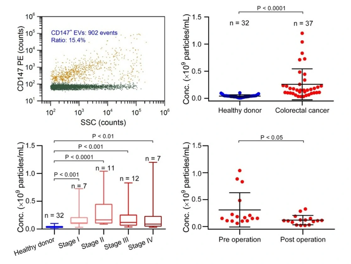
Although various methods exist to quantify EVs, EV quantification in clinical samples remains challenging and more importantly, current approaches are often unable to identify EV subpopulations. Here, we provide a nano-flow cytometry based assay for the early diagnosis of colorectal cancer.

1678329282348398.webp

Figure 1. Protein profiling of single EVs isolated from cell culture.        Figure 2. Western blotting analysis of CD147-positive EVs.
1678329287198055.webp

Figure 3. EVs for the early diagnosis of colorectal cancer.

The Flow NanoAnalyzer enables discrimination of EVs extracted from plasma of colorectal cancer patients and healthy donors after immunofluorescent labeling.


ACS Nano, 2018, 12(1), 671-680.

Nasopharyngeal Carcinoma

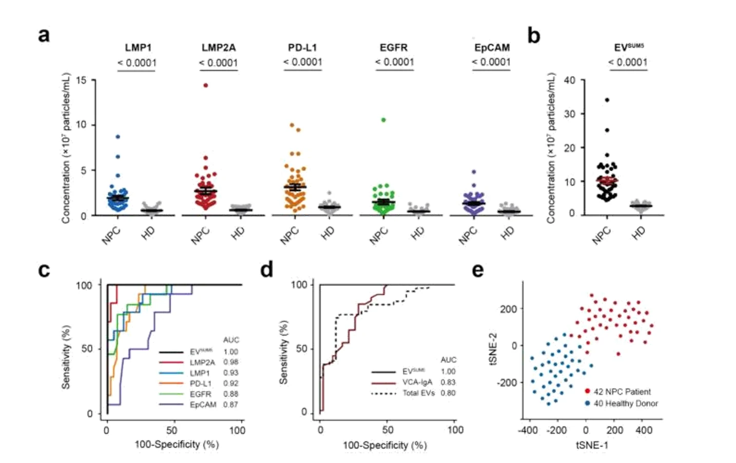
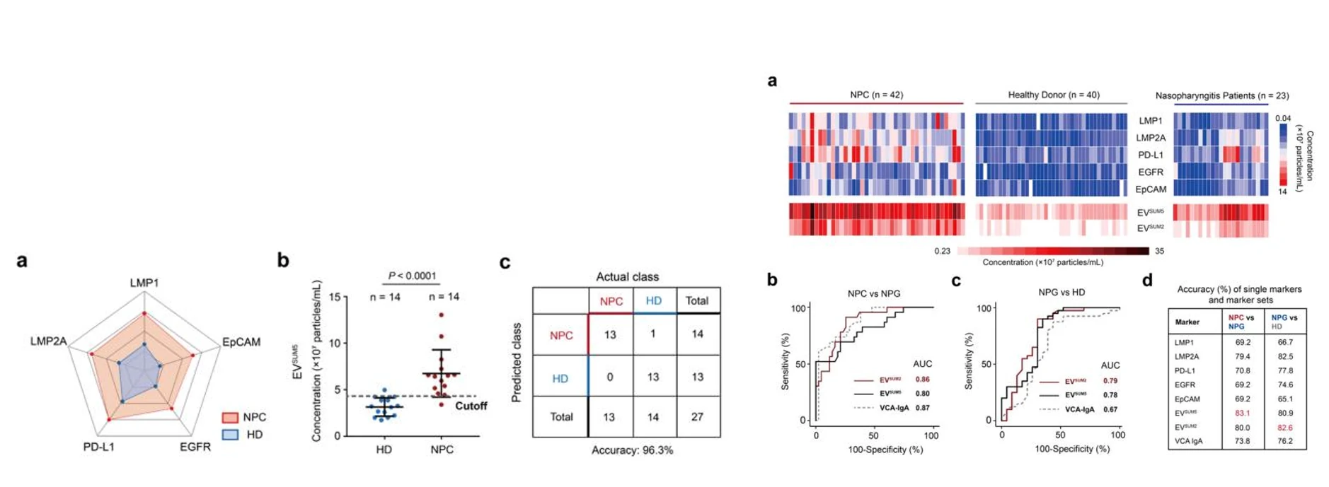
Nasopharyngeal carcinoma (NPC) is a malignant tumor commonly associated with Epstein–Barr virus (EBV) infection. Its early diagnosis as well as differentiation from nasopharyngitis (NPG) remain challenging due to the insufficient sensitivity of clinical routine screening methods. Circulating extracellular vesicles (EVs, 40–1000 nm) have shown potential in liquid biopsy for cancer diagnosis and prognosis. Herein, nano-flow cytometry (nFCM) capable of single EV analysis was applied to examine the expression of surface proteins with very low copy numbers on individual EVs as small as 40 nm. The particle concentrations of five EV subsets exposing EBV-encoded latent membrane proteins (LMP1 and LMP2A) and tumor markers (PD-L1, EGFR, and EpCAM) in plasma were determined rapidly via single-particle enumeration. The Flow NanoAnalyzer provides a rapid, reliable and non-invasive method for the diagnosis of NPC, and more accurate screening of NPC, NPG patients and healthy donors in clinical practice.

1678330084482535.webp

Figure 1. Detection of five markers of Nasopharyngeal Carcinoma

1678330092979086.webp

                           Figure 2. Diagnosis of Nasopharyngeal Carcinoma               Figure 3. Distinction of nasopharyngeal carcinoma,

                                                                                                            NPG patients and healthy donors


Anal. Chem., 2022, 94(27), 9740-9749.

Breast cancer

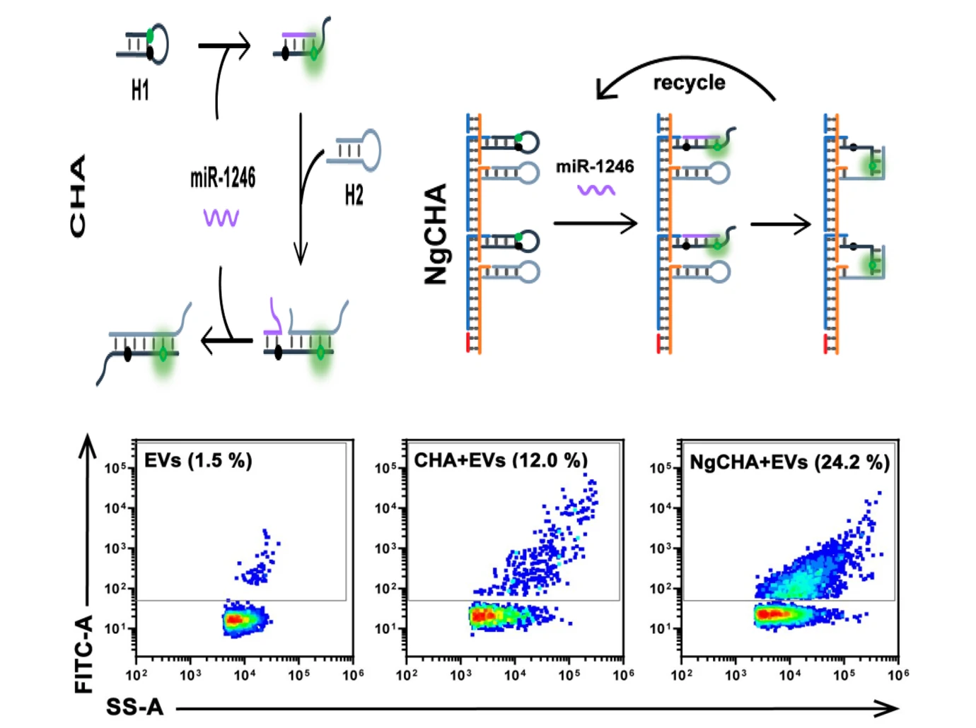
Extracellular vesicle-associated miRNAs (EV-miRNAs) are emerging as a new type of noninvasive biomarker for disease diagnosis. Their relatively low abundance, however, makes accurate detection challenging. In this article, the authors designed a DNA nanowire guided-catalyzed hairpin assembly (NgCHA) nanoprobe for profiling EV-miRNAs. NgCHA showed high penetrability to EVs, which allowed rapid delivery of the probes into EVs. In the presence of targeted miRNAs within EVs, a fluorescent signal could be generated and amplified by confining the catalytic hairpin assembly system within the nanowires, thus greatly enhancing the analytical sensitivity. In the study, co-localisation analysis of NgCHA and EVs was performed using the Flow NanoAnalyzer to confirm that NgCHA enters the EVs lumen and generates a fluorescent signal. The platform is highly sensitive, cost-saving and simple to use compared to existing methods. Therefore, it has applications for noninvasive diagnosis of cancer.

1678330494118691.webp

Figure 1. Colocalization analysis of NgCHA labeling positivity and EVs

The Flow NanoAnalyzer can achieve simultaneous detection of the scattering and fluorescence signal of the particles, i.e. co-localization analysis, with the advantage of high detection sensitivity and speed.


ACS Sensors, 2022, 7(4), 1075-1085.

Pancreatic Cancer

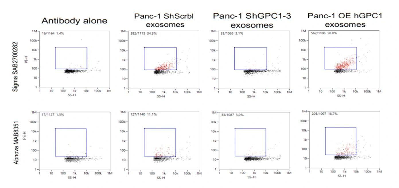
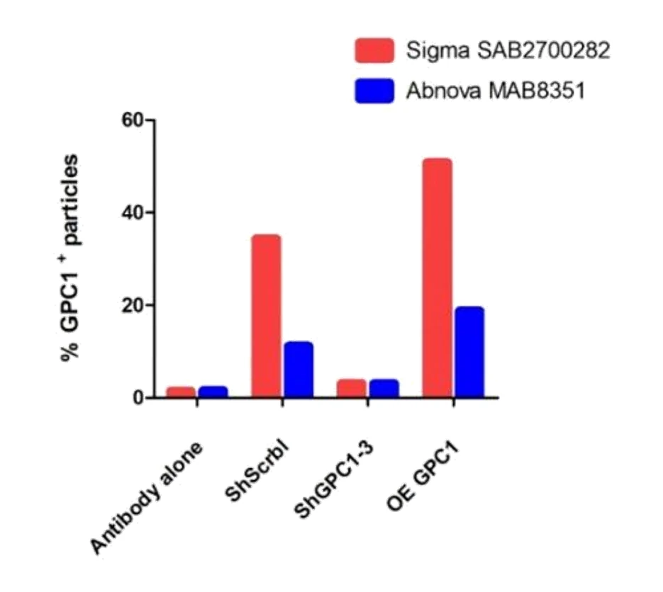
Exosomes are nano-sized vesicles shed by all cells, including cancer cells.  As they carry phospholipids, nucleic acids and proteins from their parent cells, exosomes can serve as novel liquid biopsies for diagnosis of cancer with potential prognostic value. In a? previous study, it was found that the number of Glypican-1 (GPC1) positive exosomes was significantly higher in the circulatory system of pancreatic cancer patients compared to normal subjects, and follow-up studies revealed an enrichment of GPC1 positive exosomes in breast, colorectal and oesophageal cancers, in addition to pancreatic cancer, suggesting that GPC1 is a promising marker for the early diagnosis of many cancers. In this study, the phenomenon of GPC1 overexpression in exosomes of pancreatic cancer patient origin was verified for the first time at the level of individual exosomes using multi-parameter analysis using the Flow NanoAnalyzer by immunofluorescence staining of exosomes from pancreatic cancer patients, patients with benign pancreatic disease and normal donors, and using a variety of antibodies to detect GPC1 specificity in exosomes extracted from sera of pancreatic cancer patients. A monoclonal antibody with highest specificity for GPC1 was successfully screened.

1678332051594009.webp

Figure 1. Proportion of GPC1+ positive exosomes analyzed by the Flow NanoAnalyzer.

1678332056601922.webp

Figure 2. Comparison of positive rates for different antibodies to GPC1.

The Flow NanoAnalyzer qualitatively characterises and quantifies the expression of GPC1 protein on the surface of individual exosomes by immunofluorescence staining methods, which provides a platform for diagnosis of early-stage cancer with EVs as carriers.


DOI: https://doi.org/10.1101/145706.