Six different drug-loading strategies

Author: admin     Date: 21 2 月, 2024

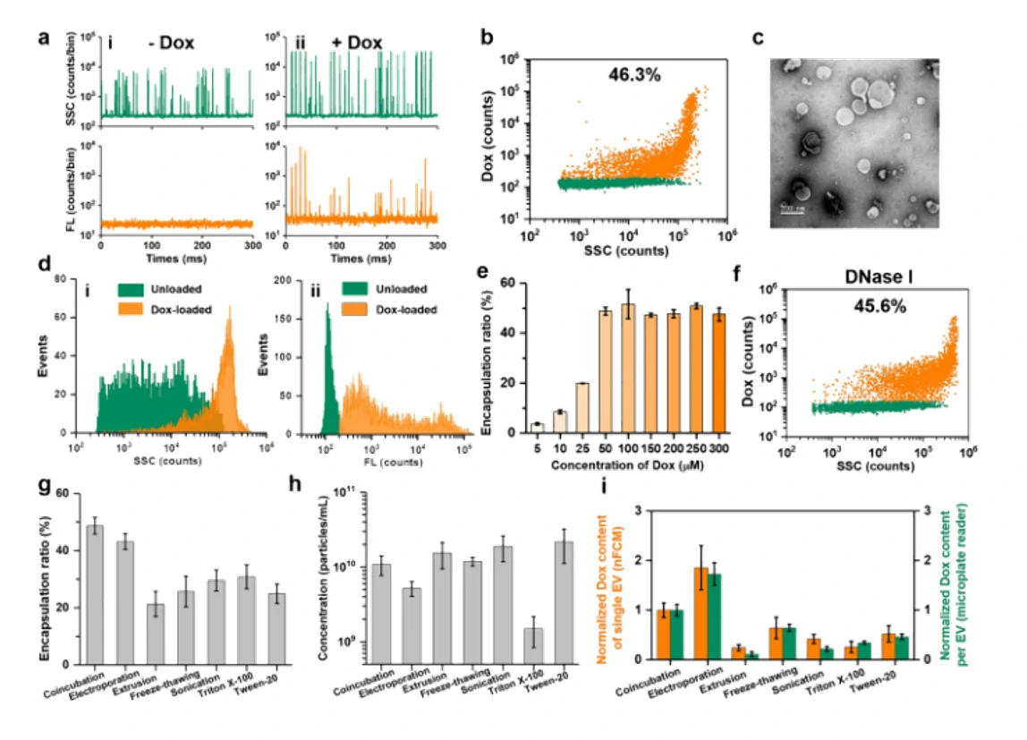
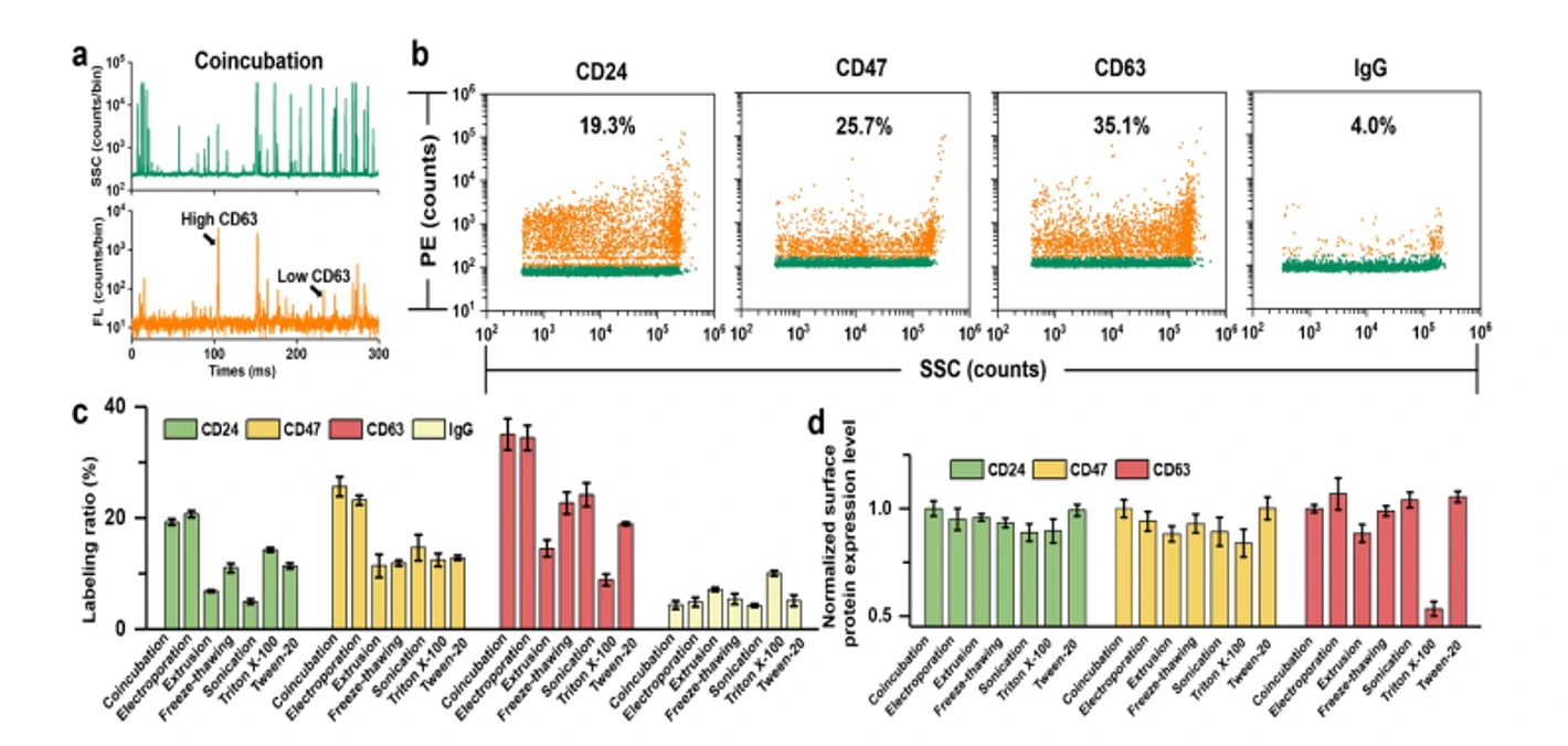
Extracellular vesicles (EVs) have emerged as an attractive drug delivery system owing to their natural roles in intercellular communication. Due to large intrinsic heterogeneity of EVs, it is highly desirable to evaluate not only the encapsulation efficiency but also the alteration of biological functionality after the drug-loading process at the single-particle level. However, the small size of EVs poses a great challenge. Taking advantage of the Flow NanoAnalyzer multiparameter analysis of single EVs as small as 40 nm, six commonly used drug-loading strategies (coincubation, electroporation, extrusion, freeze-thawing, sonication, and surfactant treatment) were explored by employing doxorubicin (Dox) as the model drug. Encapsulation ratio, EV concentration, drug content, and membrane proteins of Dox-loaded EVs were measured at the single-particle level. Data indicated that coincubation and electroporation outperformed other methods with an encapsulation ratio of approximately 45% and a higher Dox content in single EVs. Interestingly, the labeling ratios of membrane proteins indicated that varying degrees of damage to the surface proteins of EVs occurred upon extrusion, freeze-thawing, sonication, and surfactant treatment. 
1678332460582687.webp

Figure 1. Encapsulation ratio of Dox-loaded EVs in different drug-loading strategies
1678332465544256.webp

Figure 2. The expression of  membrane proteins of Dox-loaded EVs in differemt drug-loading strategies 

Confocal fluorescence microscopy and nano-flow cytometry analysis revealed that Dox-loaded EVs prepared by electroporation induced the strongest apoptosis followed by coincubation. These results correlated well with their cellular uptake rate and fundamentally with the Dox encapsulation efficiency of single EVs. Flow The NanoAnalyzer provides a rapid and sensitive platform for single-particle assessment of drug-loading strategies for incorporating drugs into EVs.


Anal Bioanal Chem., 2022. https://doi.org/10.1007/s00216-022-04248-4.

EVs isolation from raw milk

Author: admin     Date: 21 2 月, 2024

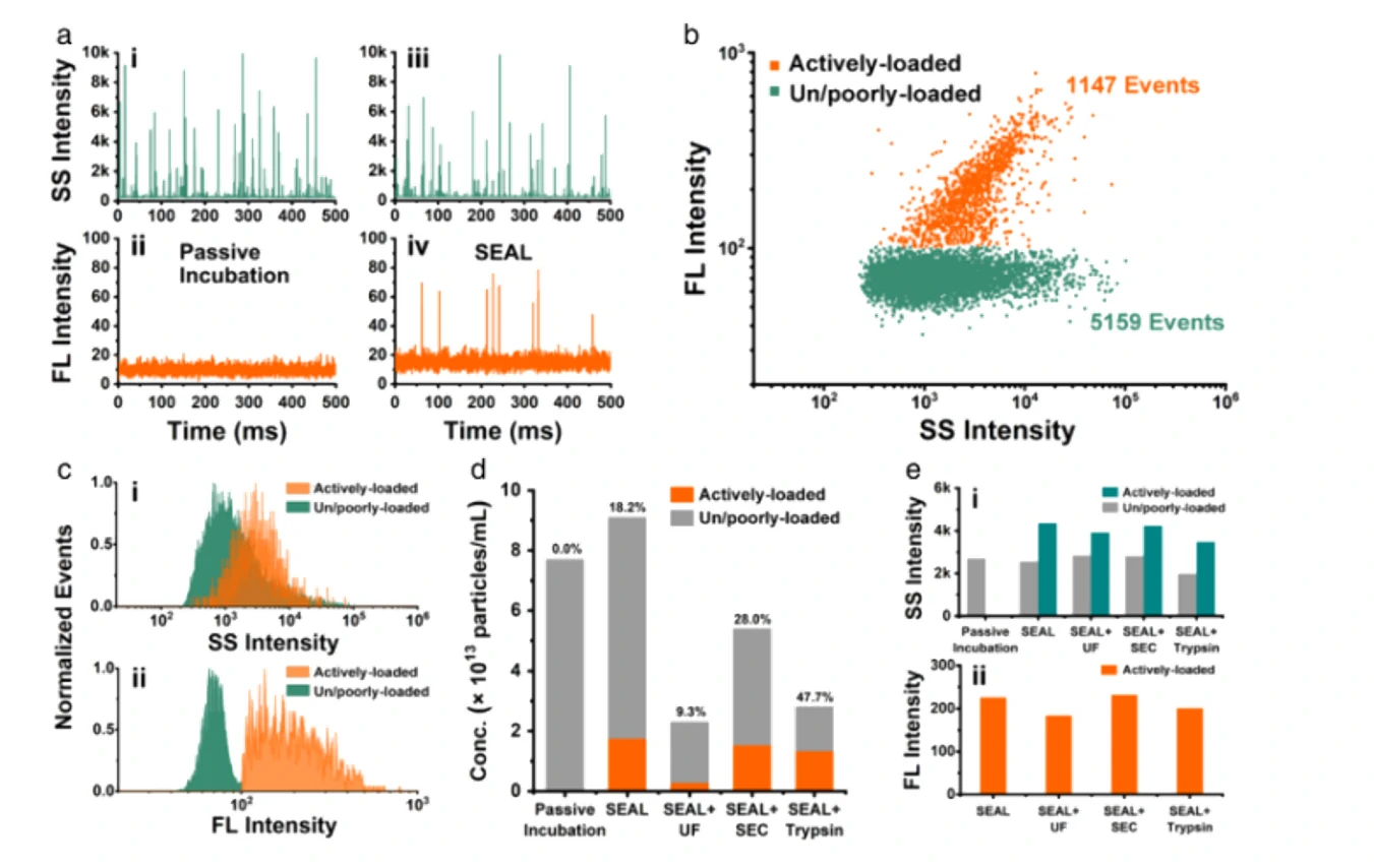
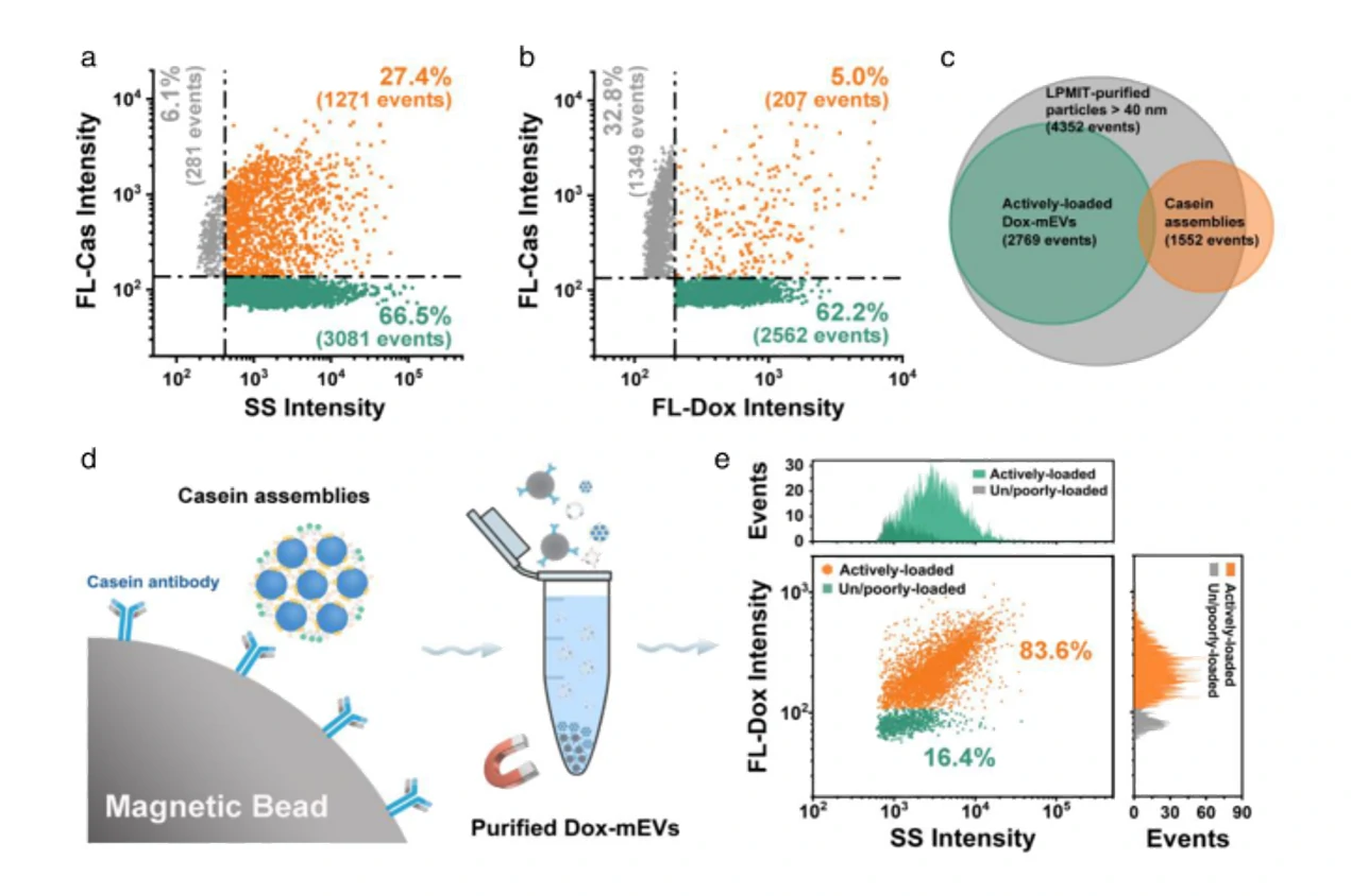
Extracellular vesicles (EVs) have demonstrated unique advantages in serving as nanocarriers for drug delivery, yet the cargo encapsulation efficiency is far from ideal, especially for hydrophilic chemotherapeutic drugs. Besides, the intrinsic heterogeneity of EVs renders it difficult to evaluate drug encapsulation behaviour. Inspired by the active drug loading strategy of liposomal nanomedicines, here we report the development of a method, named “Sonication and Extrusion-assisted Active Loading” (SEAL), for effective and stable drug encapsulation of EVs. Using doxorubicin-loaded milk-derived EVs (Dox-mEVs) as the model system, sonication was applied to temporarily permeabilize the membrane, facilitating the influx of ammonium sulfate solution into the lumen to establish the transmembrane ion gradient essential for active loading. Along with extrusion to downsize large mEVs, homogenize particle size and reshape the nonspherical or multilamellar vesicles, SEAL showed around 10-fold enhancement of drug encapsulation efficiency compared with passive loading. Single-particle analysis by the Flow NanoAnalyzer was further employed to reveal the heterogeneous encapsulation behaviour of Dox-mEVs which would otherwise be overlooked by bulk-based approaches. Correlation analysis between doxorubicin auto-fluorescence and the fluorescence of a lipophilic dye DiD suggested that only the lipid-enclosed particles were actively loadable. Meanwhile, immunofluorescence analysis revealed that more than 85% of the casein positive particles was doxorubicin free. These findings further inspired the development of the lipid-probe- and immuno-mediated magnetic isolation techniques to selectively remove the contaminants of non-lipid enclosed particles and casein assemblies, respectively. Finally, the intracellular assessments confirmed the superior performance of SEAL-prepared mEV formulations, and demonstrated the impact of encapsulation heterogeneity on therapeutic outcome. 

1678332729273995.webp

Figure 1.Single particle analysis of the drug loading capacity of SEAL-prepared Dox-mEVs by nano-flow cytometry. 

1678332734311344.webp

Figure 2. Multi-parameter analysis of the correlation between drug loading capacity and lipid labelling pattern of Dox-mEVs. 

The developed cargo-loading approach and the Flow NanoAnalyzer-based characterization method provides instructive insight into the development of EV-based delivery systems.


J Extracell Vesicles, 2021, 10:e12163.

Red blood cell-derived EVs in AML therapy

Author: admin     Date: 21 2 月, 2024

Acute Myeloid Leukaemia (AML) is the most common blood cancer in adults. Although 2 out of 3 AML patients go into total remission after chemotherapy and targeted therapies, the disease recurs in 60%-65% of younger adult patients within 3 years after diagnosis with a dramatically decreased survival rate. Therapeutic oligonucleotides are promising treatments under development for AML as they can be designed to silence oncogenes with high specificity and flexibility. However, validated approaches for safely and efficiently delivering oligonucleotide drugs are limited. This issue could be resolved by utilizing a new generation of delivery vehicles such as extracellular vesicles (EVs).

A platform to purify large-scale quantities of red blood cell-derived EVs (RBCEVs) (1013–1014 EVs from each blood unit) in a low cost and time-efficient fashion has been validated. In a previous study, a variety of RNA therapeutics including ASOs, gRNAs, and Cas9 mRNA were loaded into RBCEVs and subsequently delivered to target cells, showing significant therapeutic effects in, in vitro and in vivo models of breast cancer and AML. In this study, the authors harness RBCEVs and engineer them via exogenous drug loading and surface functionalization to develop an efficient drug delivery system for AML and characterise the single particles using the Flow NanoAnalyzer.

1678333263362567.webp

                                                   Figure 1. EVs surface antibody modification strategies                         Figure 2. The Flow NanoAnalyzer provides an estimation of the modification efficiency

To estimate the antibody conjugation efficiency at single-RBCEV level, the Flow NanoAnalyzer was used to detect RBCEVs coated with biotinylated anti-CD33 antibody and isotype monoclonal antibody (IgG-EV). The nano-flow cytometric analysis indicated that ~74% of RBCEVs were successfully conjugated with the CD33 monoclonal antibody. Subsequent in vivo experiments demonstrated that RBCEVs loaded with ASO significantly suppresses AML progression. Nano-flow cytometry plays a crucial role in the validation of exosome modification results and is expected to advance the development of engineered EVs drugs.


Cell Prolif., 2022, 55(9):e13255.

Antibody Drug

Author: admin     Date: 21 2 月, 2024

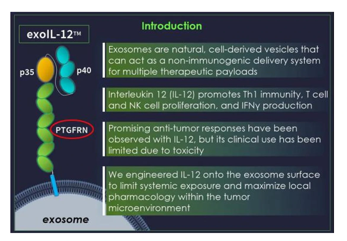
ExoIL-12 from Codiak BioSciences is the first engineered clinically exosome therapeutic candidate in the world. Codiak obtained high purity exosomes by density gradient centrifugation, and used proteomic analyses on purified EVs, identifying two protein families previously unexplored as EV scaffolds, the EWI immunoglobulin superfamily (IGSF8 and PTGFRN) and the MARCKS protein family (MARCKS, MARKCSL1, and BASP1). Members of both protein families were found to be abundant in EVs derived from a variety of cell types and were selected for further investigation as scaffold proteins for EV loading. These scaffold proteins can be genetically modified to attach various biomolecules or drugs, such as cytokines, antibody fragments, RNA binding proteins, vaccine antigens, etc., which makes them a ideal platform for drug delivery. 

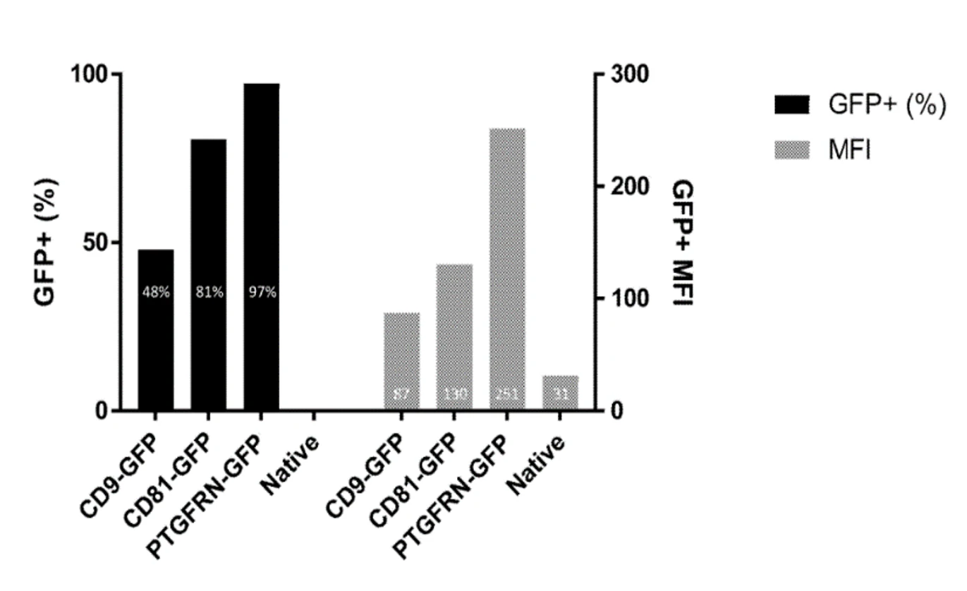
Researchers assessed the relative ability of these proteins to direct fusion partners into EVs using FLAG-tagged GFP as a surrogate cargo molecule and have attempted to quantitatively analyse them using traditional flow cytometery and ELISA methods. However, the traditional cytometry only qualitatively demonstrate that these proteins are expressed at the cellular level, cannot prove whether the target protein is effectively packaged into EVs. Meanwhile, ELISA can only determine that a protein is overexpressed but not whether this overexpression is prevalent in EVs or abounds in a subpopulation. Therefore, it is necessary to analyse EVs at the single particle level. Using the Flow NanoAnalzyer to identify scaffold proteins, and analyze the expression of several scaffold proteins at single EVs particle level, the researchers showed that PTGFRN protein was expressed in 97% of exosomes. In contrast to the semi-quantitative Western Blot and the ELISA method which can only provide an average value, the Flow NanoAnalzyer can determine the proportion of positive subpopulations. Furthermore, the fluorescence intensity can be used to further reflect information on the amount of protein expression on single EVs particle.

1678340062373154.webp

Figure 1. The expression of PTGFRN proteins and IL2

1678340094487970.webp

Figure 2. Proteins’s different proportion of expression on the Flow NanoAnalyzer

The application of the Flow NanoAnalzyer is the first single particle assay for genetically modified PTGFRN exosomes at single particle level. Traditional ELISA can only quantify the protein on a large number of exosomes, while the Flow NanoAnalzyer can identify and quantify the protein expression at the individual exosome level, and found that PTGFRN is an ideal scaffold protein with expression percentage of 97%.

Molecular Therapy, 2021, 29(5), 1729-1743.

Nucleic acid drugs

Author: admin     Date: 21 2 月, 2024

It has been demonstrated recently that extracellular vesicles (EVs) carry DNA; however, many fundamental features of DNA in EVs (EV-DNA) remain elusive. In this study, a laboratory-built nano-flow cytometer (nFCM) that can detect single EVs as small as 40 nm in diameter and single DNA fragments of 200 bp upon SYTO™ 16 staining was used to study EV-DNA at the single-vesicle level. Through simultaneous side-scatter and fluorescence (FL) detection of single particles and with the combination of enzymatic treatment, present study revealed that: (1) naked DNA or DNA associated with non-vesicular entities is abundantly presented in EV samples prepared from cell culture medium by ultracentrifugation; (2) the quantity of EV-DNA in individual EVs exhibits large heterogeneity and the population of DNA positive (DNA+ ) EVs varies from 30% to 80% depending on the cell type; (3) external EV-DNA is mainly localized on relatively small size EVs (e.g. <100 nm for HCT-15 cell line) and the secretion of external DNA+ EVs can be significantly reduced by exosome secretion pathway inhibition; (4) internal EV-DNA is mainly packaged inside the lumen of relatively large EVs (e.g. 80-200 nm for HCT-15 cell line); (5) double-stranded DNA (dsDNA) is the predominant form of both the external and internal EV-DNA; (6) histones (H3) are not found in EVs, and EV-DNA is not associated with histone proteins and (7) genotoxic drug induces an enhanced release of DNA+ EVs, and the number of both external DNA+ EVs and internal DNA+ EVs as well as the DNA content in single EVs increase significantly. 

微信截图_20230901162541.webp


Figure 1. Characterization of DNA in EVs (EV-DNA)                  

微信截图_20230901162752.webp

Figure 2. Identification of nucleic acids in EVs

1678340530482520.webp

Figure 3. The impact of exosome secretion inhibitor GW4869 on EV-DNA.

Size exclusion chromatography (SEC) was found to be ineffective in separating free DNA from EVs, while density gradient centrifugation removed the vast majority of free DNA. This study provides direct and conclusive experimental evidence for an in-depth understanding of how DNA is associated with EVs.


J Extracell Vesicles, 2022, 11:e12206.

EV-inspired liposomes

Author: admin     Date: 21 2 月, 2024

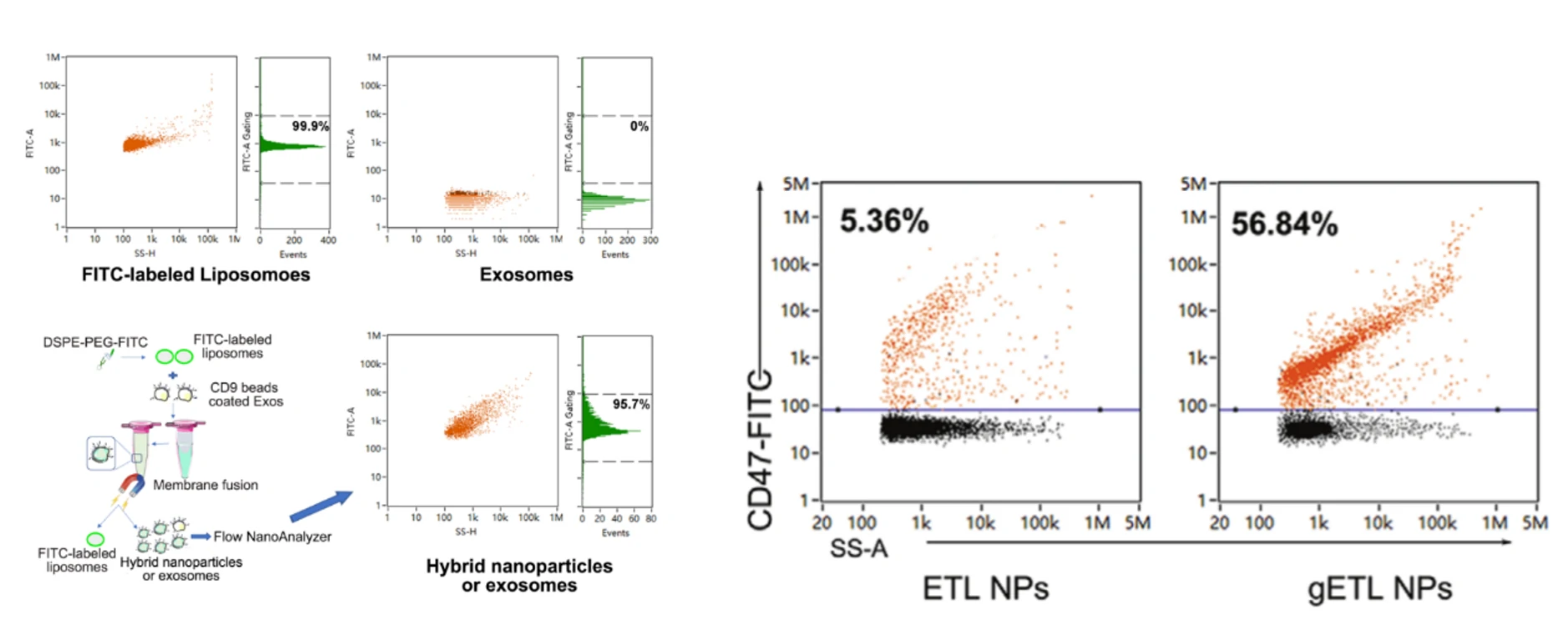
EVs can be genetically engineered to overexpress CD47, a protein that enables cargos to escape from clearance by the immune system and lead to enhanced accumulation. Liposomes are synethetic nanoparticles that capable of encapsulating thermosensitive drugs. The fusion of these two nanoparticles can exert the advantages of both exosomes and liposomes, with an effect of “1 + 1 > 2”. The resulting genetically engineered exosomes-thermosensitive liposomes hybrid nanoparticles (gETL NPs) bears CD47 proteins on the surface and thermosensitive drugs inside. The Flow NanoAnalyzer was employed to analyze the liposomes, exosomes and gETL NPs at single-particle level, which played important roles in the optimization and screening of conditions for genetically engineered exosomes and the production of gETL NPs.

1676439213802119.webp

1678341065876070.webp

                             Figure 1. Exosomes-Liposomes hybrid nanoparticles                    Figure 2. Detection of CD47 expression ratio in exosome-liposome nanoparticles


Adv. Sci., 2020, 7(18), 2000515.