Comparison of Commercial Isolation kits

Extracellular vesicles (EVs) are nanosized particles with significantly varied sizes. In addition, EVs are also heterogeneous in composition, and enriched in membrane-associated, high-order oligomeric protein complexes. EVs are being developed as diagnostic and therapeutic agents in multiple disease models. Isolation of EVs from complex biological fluids with high purity is essential to the accurate analysis of EV cargo. Unfortunately, commonly used isolation techniques do not offer good separation of EVs from non-EV contaminants. Hence, it is important to have a standardized method to characterize the properties of EV preparations, including size distribution, particle concentration, purity and phenotype. The current common methods of exosomes purification include; differential ultracentrifugation, size exclusion, exosome precipitation and immunoaffinity. Characterization of EVs single particle level is of great importance to investigate the mechanism of vesicle transport systems, cell communication, early diagnosis, and disease treatment. The current techniques for single particle detection include cryoTEM, nanoparticle tracking analysis (NTA), tunable resistive pulse sensing (TRPS). However, with exception to cryoTEM, the other techniques struggle to detect particles smaller than 70 nm and only provide size distribution and particle concentration. 

The Flow NanoAnalyzer enables multiparameter analysis of single EVs as small as 40 nm, a new benchmark for the quality and efficiency assessment of EVs isolated from plasma is reported. Properties, such as size distribution, particle concentration, purity, recovery rate and surface proteins, were measured. The performance of five widely used commercial isolation kits was examined and compared with the commonly used technique of differential ultracentrifugation.

 1678176783167626.webp

Figure 1. The particle size distribution and concentration                       Figure 2. Particle concentration of 

     of EVs preparation from PFP by different methods.                             EV preparations from PFP and VD-PFP.

1678176789264882.webp
          Figure 3. Measurement of the purity of different                              Figure 4. Measurement of the recovery rates of  

 isolation methods for pure EVs.                                                      different isolation methods for pure EVs.

1678176796619996.webp

Figure 5. Single-particle phenotyping of EVs isolated by UC from CCCM of HCT15 cells and PFP.

The purity of EVs isolated from PFP by UC was 78.2% in this study, which is only slightly lower than the 90% for EVs isolated from conditioned cell culture medium. In contrast to the much higher yields of commercial isolation kits, the measured purity (ranging from 28.1 to 5.3%) was markedly lower in comparison to UC.

To compare characterization of surface proteins on EVs, the EVs extracted by the kit also need to be further removed by the ultra-ionization method to obtain results consistent with UC. In conclusion, there is a significant difference between commonly used isolation kits and traditional ultracentrifugation methods.

J Extracell Vesicles, 2019, 9(1), 1697028.

Evaluation of purification methods of EVs

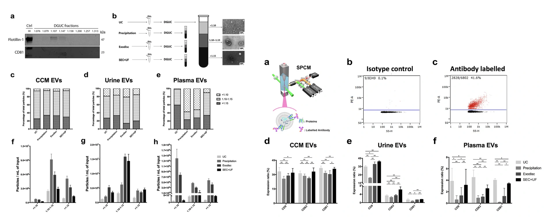
One of the challenges that restricts the growth of extracellular vesicle (EV) research field is the lack of a standardized method for EV separation. In this study, three commonly used EV separation methods, ultracentrifugation, precipitation and size exclusion chromatography (combined with ultrafiltration with a microfluidic tangential flow filtration device, Exodisc) was used to comprehensively evaluate the EV yield and sample purity from cell culture medium, human urine and plasma.

The isolated EVs were assessed in terms of their yield, purity and surface protein expression by the Flow NanoAnalyzer. Using these parameter, practical recommendations were made for the choice of isolation and purification methods based on sample type and downstream applications. 

1678172976589435.webp

                         Figure 1. Concentrations of particles separated by different methods                       Figure2.  Purity of EV samples assessed by

                                                                                                                                        particle/protein ratio


1678173000844346.webp

Figure 3. Composition purity analysis of EVs by DGUC           Figure 4. Purity of EV samples assessed by single particle phenotyping

1678173022581027.webp

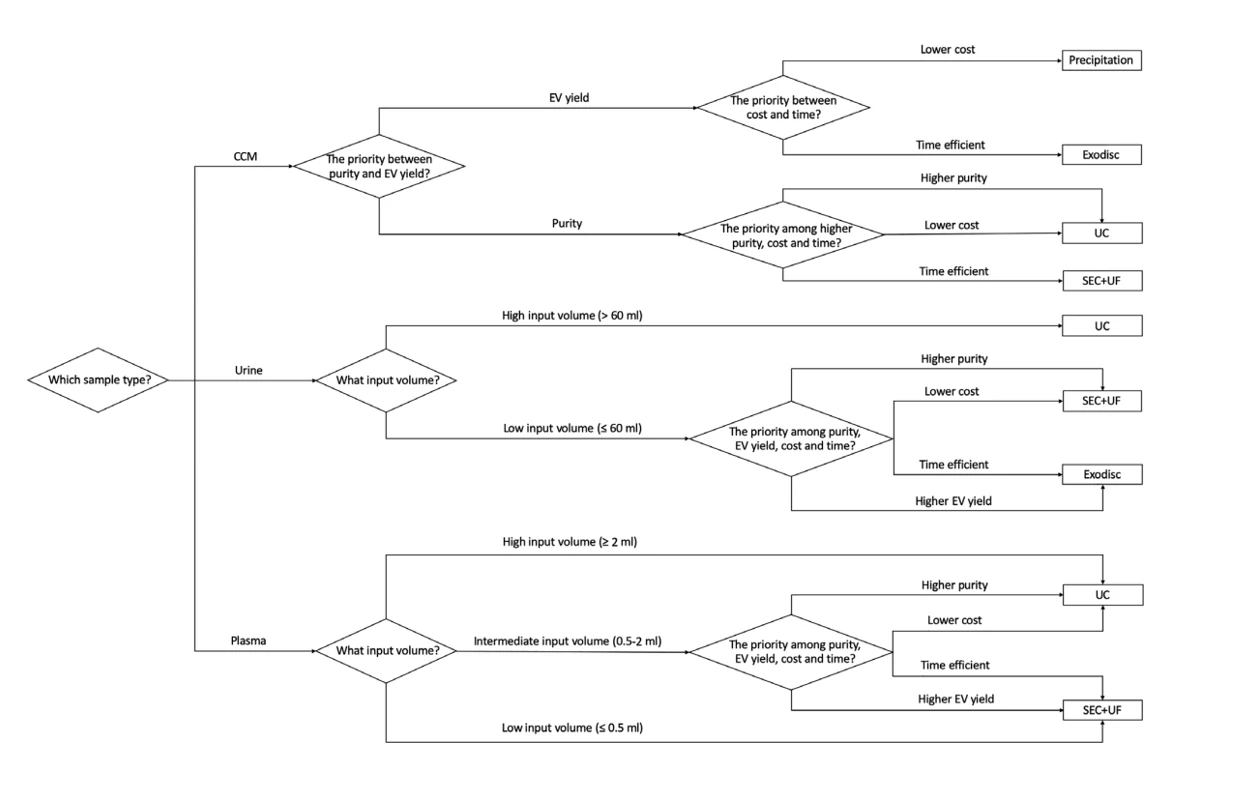
Figure 5. The decision tree on how to select the proper EV separation method based on the input sample


In this study, the author characterized EVs by the Flow NanoAnalyzer with multiple parameters, evaluated purification methods suitable for different sample types, concluded how to select suitable purification methods based on different sample sources and downstream applications, and provided practical recommendations.

J Extracell Vesicles, 2020, 10(2):e12044.

Optimization of clinical isolation of EVs

To acquire high-quality EVs from complex samples like plasma, a simplified dichotomic SEC method was established. Utilizing the Flow NanoAnalyzer, particle concentration and size distribution was first determined. The  purity was assessed by implementing CFSE labelling of EVs. The dichotomic SEC method was found to separate particles in serum or plasma into two components: EVs and non-vesicular contaminates. Corroborating this, proteomics showed that the dichotomic SEC method can remove different co-isolating contaminant proteins from human plasma with comparable purity to that of UC. Collectively, this isolation method shows an intriguing potential in the preparation of EVs toward clinical testing and/or basic research.

1678175341541421.webp

Figure 1. Optimization of SEC for plasma EV isolation

By implementing the multi-analysis capabilities of the Flow NanoAnalyzer it was found that the dichotomic SEC and ultracentrifugation could isolate EVs from human plasma with comparable purity and even higher extraction rate by SEC.

J Extracell Vesicles, 2021, 10:e12145.

Isolation of native EVs from primary biofluids

The isolation and purification of EVs from body fluids is challenging due to the complex composition of the fluids themselves. Complex fluids include; blood, urine , ascites and so on. The particles found in the fluid can have an overlap in terms of size and composition with EVs, resulting in high sample impurity. The most used method of EV extraction, to obtain high purity EVs from complex biofluids, include differential centrifugation and density gradient centrifugation. The method has several disadvantages, such as being time-consuming, complicated, as well as resulting in low particle yield and quality due to excessive centrifugal force method, which can affect the integrity and composition of EVs. These disadvantages restrict its translation to clinical and industrial applications.

1678177963885639.webp

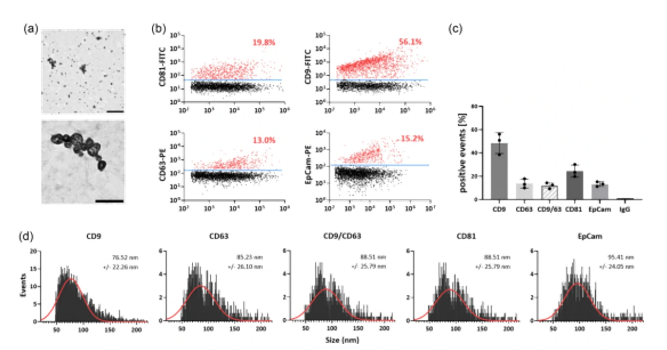
Figure 1.  Single particle characterization of EVs via electron microscopy and NanoFCM 

Researchers have optimized the FFE purification method via combining the downstream analysis methods; western blot, TEM and nano-flow cytometry. This has resulted in the acquisition of isolated and purified EVs from highly complex body fluid in a fast and high-throughput manner.  

Journal of Extracellular Biology, 2022, 1, e71.