Ag@sio2 Core-Shell Nanoparticles
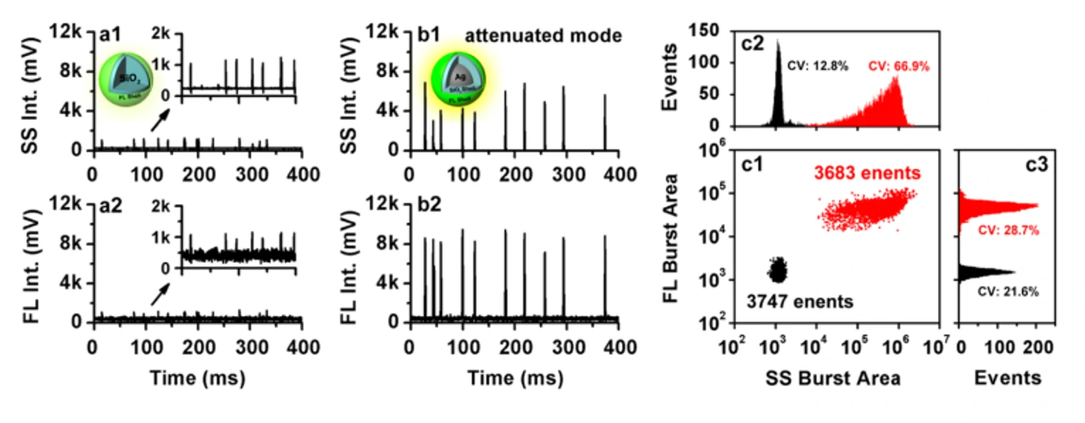
Metal-enhanced fluorescence (MEF) based on localized surface plasmon resonance (LSPR) is an effective method to improve the sensitivity of detection. Plasmonic nanoparticles are inherently heterogeneous, so single-particle analysis of MEF in free solution is key to understanding and controlling the MEF process. In this study, the Flow NanoAnalyzer was used to study the fluorescence enhancement of a single plasmonic nanoparticle near to fluorophore molecules. The study used Ag@SiO2 core-shell nanoparticles as a model system, which consist of a silver core, a silica shell, and a thin layer of FITC-doped silica shell. FITC-doped silica nanoparticles of the same size but with no silver nucleus as their counterparts were used. The Flow NanoAnalyzer was employed to detect side-scattering and fluorescence signals of single particles in the suspension and systematically study the effects of the size of silver nuclei (40-100 nm) and the distance between fluorophore and metal (5-30 nm).

Figure 1. Analysis of MEFs at the Single Particle Level by the Flow NanoAnalyzer

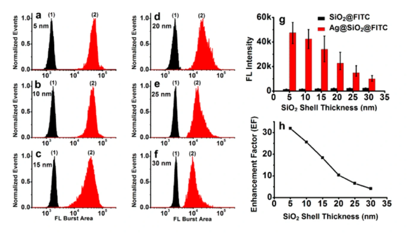
Figure 2. Analysis of Ag@SiO2 core-shell nanoparticles with a silver core size of 70 nm and different silica shell thicknesses by the Flow NanoAnalyzer
The experimental observations in this study at the single-particle level are well supported by finite-difference time-domain (FDTD) calculations compared with ensemble-averaged fluorescence spectral measurements. This is very important for the design and control of plasmonic nanostructures, which can effectively enhance the fluorescence.
ACS Sens., 2017, 2(9), 1369-1376.


