Measurement of complete lentivirus particles

Lentivirus is a gene vector developed from HIV-1 (human immunodeficiency type I virus), composed of a lipid membrane, capsid, and internal RNA genome with an average diameter of 80-120 nm. Lentiviral vectors now play a critical role in the field of cell and gene therapies, especially chimeric antigen T-cell (CAR-T) therapy. Currently, the most frequently used methods for titration of lentivirus vectors are:

1) Transducing Units (TU): Based on the capability of transducing a cell and expressing the transgene.

2) Copy Number Determination: Based on real-time quantitative PCR (qPCR).

3)Analysis of Viral p24 Concentration: Using enzyme-linked immunosorbent assay (ELISA).

Each method provides separate important data about a lentiviral sample. Nano-flow cytometry can be employed to describe all these lentiviral characteristics simultaneously at a single particle level. Additionally, the use of the Flow NanoAnalyzer on lentiviral products identifies both mature lentiviruses and contaminant unincorporated components.

Multiple components coexist in lentiviral samples, such as mature virions, partially assembled virions, free nucleic acids, etc. Analysis by nano-flow cytometry can give a thorough description of the active population of lentivirus presenting VSV-G for cellular recognition and infectivity, and internal nucleic acid cargo for activity. This data is highly quantitative, achieved rapidly, and simultaneously measures the other immature particles to determine product purity.
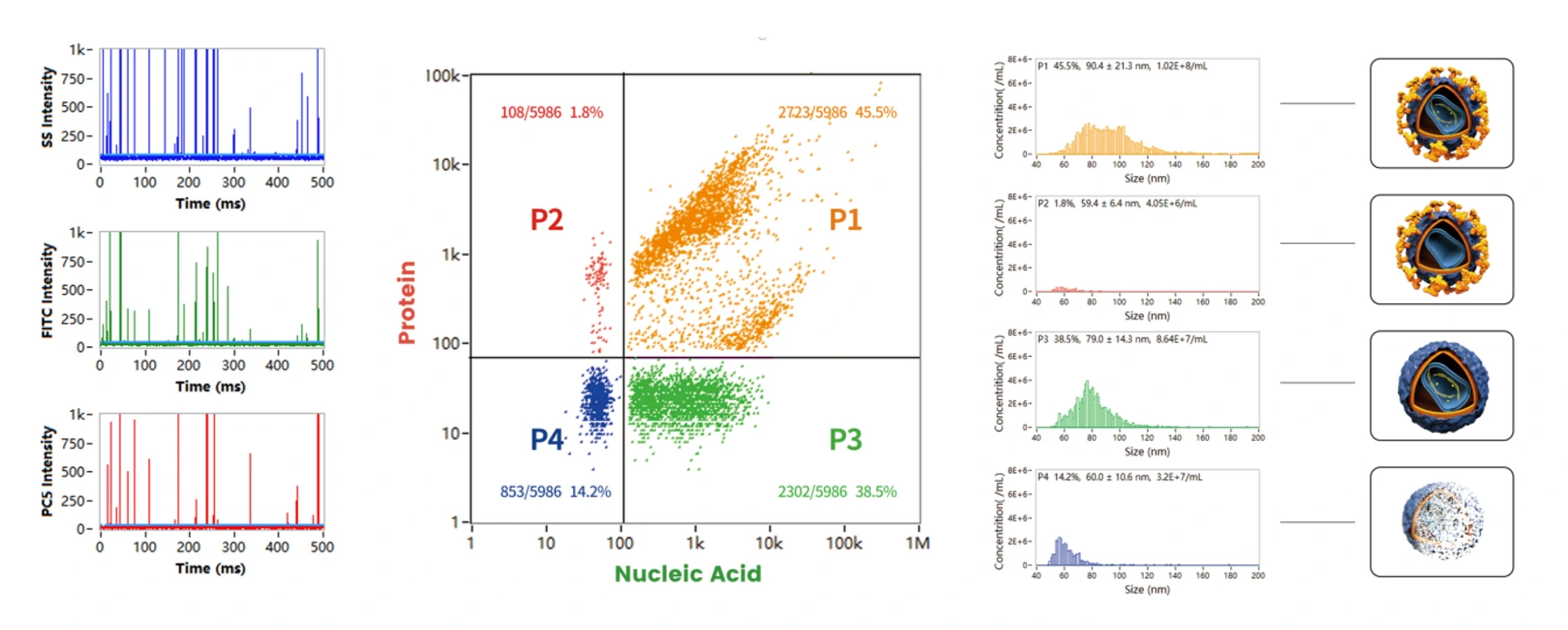
1678343978587375.webp

Figure 1. Analyze different compositions of lentivirus particles on the Flow NanoAnalyzer

In Figure 1, the P1 population represents the complete lentivirus, double-positive for labeled nucleic acid and VSV-G proteins, making up ~45% of the particle product, with additional size distribution and concentration data provided in a single 2-3 minute run time. The major particle contaminant is the VSV-G-negative, genome-positive population. Despite the potential for these VSV-G-negative particles to be insufficient for gene integration, they could skew qPCR measurements and particle counting methods.


Copyright © 2022 NanoFCM Inc.

Extracellular Vesicles Encapsulated Oncolytic Viruses for the Treatment of Tumor Diseases

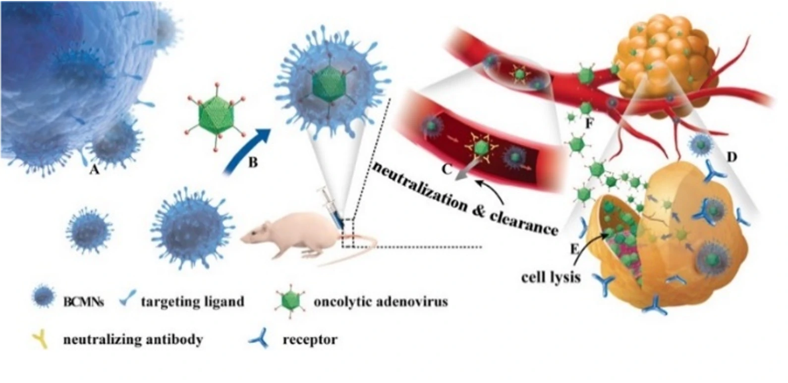
Extracellular vesicles possess the ability to evade the immune system and traverse the blood-brain barrier. They can also target tumor cells through specific markers, making them one of the most ideal carriers for drug treatment of tumor diseases. Oncolytic viruses (OAs) are tumor-killing viruses with replication ability, selectively infecting tumor cells by deactivating tumor suppressor genes in target cells, replicating in their cytoplasm, and eventually destroying them. They also stimulate the immune response, attracting more immune cells to continue killing residual cancer cells. However, due to the innate and antiviral immunity of the human body, oncolytic adenoviruses cannot reach specific tumor tissues, limiting their application in tumor treatment.

This study engineered extracellular vesicles with targeting functions and antiviral immunity, including the encapsulation of oncolytic adenovirus into extracellular vesicles. Exosome-encapsulated oncolytic adenovirus can resist the innate and acquired immunity of the human body. It can specifically infect tumor cells, self-replicate to form a large number of viruses that specifically target tumors, and subsequently kill tumor cells.

1676450490162794.webp

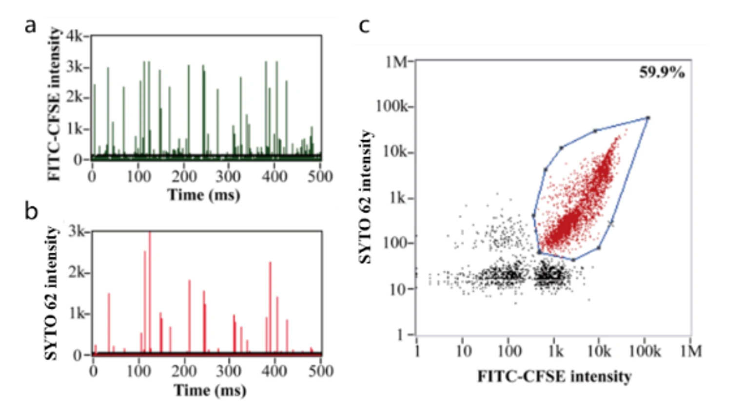
This study demonstrates that exosome-encapsulated oncolytic viruses exhibit antiviral immunity, and the targeting of tumor cells can be enhanced by labeling tumor-specific markers on the surface of exosomes. Releasing the oncolytic adenovirus  into the exosomes is a key step in the experiment. The encapsulation efficiency and effectiveness of oncolytic adenovirus directly impacts the subsequent treatment outcomes. The researchers used fluorescent dye CFSE and nucleic acid dye SYTO™ 62 to label exosomes and OA, respectively. The Flow NanoAnalyzer was employed to evaluate the encapsulation efficiency of oncolytic adenovirus.

1678345208870857.webp

Figure 1. Encapsulation efficiency of oncolytic viruses.

Based on nucleic acid staining, the Flow NanoAnalyzer allows the determination of the encapsulation efficiency of oncolytic adenoviruses, which was 59.9% in this case.


Nano Lett., 2019, 19(5), 2993-3001.

Quantitative Assessment of the Physical Virus Titer and Purity

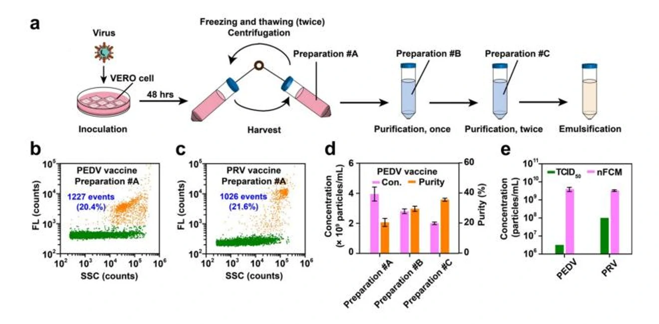
Rapid quantification of viruses is vital for basic research on viral diseases as well as the clinical application of virus-based products. The plaque assay is the classical method used for virus quantification, yet it requires 1-2 weeks (for mammalian viruses) or 1-2 days (for bacteriophages) to determine the infectious titers. Although the enzyme-linked immunosorbent assay (ELISA) and quantitative polymerase chain reaction (qPCR) can quantitatively measure specific proteins and the nucleic acid sequences of viruses, respectively, the stringency of these "bulk" analytical methods is low. They are incapable of distinguishing proteins originating from complete virions or empty capsids, or soluble viral proteins and viral nucleic acids packed inside the capsid or that are free in solution.

1678344903661801.webp

Figure 1. Characterization of virus-based vaccines.

A high-throughput single-particle method to enumerate intact viral particles using the Flow NanoAnalyzer was reported. Using the bacteriophage T7 as a model system, intact virions were completely discriminated from empty capsids and naked viral genomes. Successful measurement of the physical virus titer and purity was demonstrated for recombinant adenoviruses, which could be used for gene delivery, therapeutic products derived from phage cocktails, and infected cell supernatants for veterinary vaccine production.


Angew. Chem. Int. Ed., 2021, 60, 9351-9356.

Drug Dlivery-Virus-like Particles

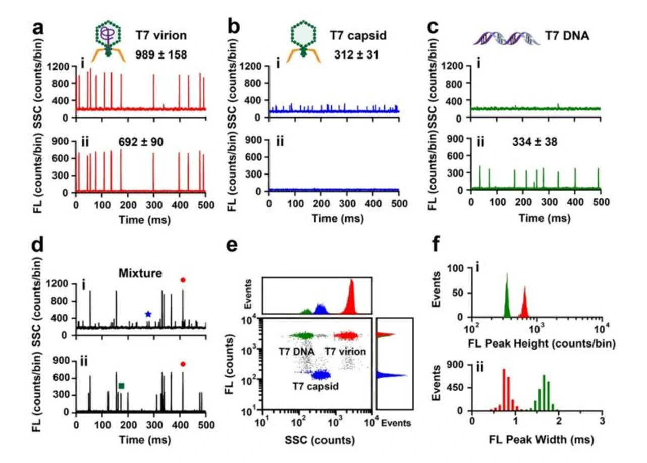
Virus-like particles (VLPs) are considered natural nucleic acid nanomedicines, in which nucleic acids (drugs) are encapsulated within a viral empty shell (drug carrier). Through nucleic acid staining, we can rapidly distinguish complete viruses, viral empty shells, free nucleic acids, etc., from the compounds. This allows us to further assess drug encapsulation efficiency, quantity, and the proportion of effective drugs.

The nucleic acid dye SYTO™ 82 was used to stain the viral (or vector) genome, and a laboratory-built nano-flow cytometer (nFCM) was employed to simultaneously detect the side-scatter and fluorescence signals of individual viral particles. Using the bacteriophage T7 as a model system, intact virions were completely discriminated from empty capsids and naked viral genomes. Successful measurement of the physical virus titer and purity was demonstrated for recombinant adenoviruses, which could be used for gene delivery, therapeutic products derived from phage cocktails, and infected cell supernatants for veterinary vaccine production.

1678345549206166.webp

Figure 1. Differentiation of intact virions, empty capsids, and free viral DNA.


Angew. Chem. Int. Ed., 2021, 60, 9351-9356.